Metal Artifact Reduction Software Offers Additional Diagnostic Advantages.
An aging population along with a wide variety of orthopaedic conditions and injuries among people of all ages are contributing to an increase in orthopaedic imaging. Although conventional multi-detector computed tomography (MDCT) offers important advantages by providing 3D imaging, specialists also need access to high-resolution visualization of anatomy in the presence of metal in weight-bearing positions.
Since traditional MDCT imaging has difficulty addressing these needs, new imaging technologies such as cone beam CT (CBCT) are being introduced. The CARESTREAM OnSight 3D Extremity System is a CBCT system that has the ability to capture and reconstruct a 3D image of upper and lower extremities. The system has a donut-shaped bore where patients insert the injured extremity. A small door allows patients to easily step into the bore for weight-bearing exams of feet, ankles and knees. Acquisition takes only 25 seconds and image reconstruction takes about five minutes. Since only the affected body part is being imaged, the dose to the patient is significantly less than traditional MDCT and all parameters are optimized exclusively for extremity imaging rather than designed as a general-purpose system.
With this new system, the detector and source rotate around the patient’s anatomy, acquiring a multitude of projections from different angles, both axially and rotationally. Each of the system’s three X-ray sources fire sequentially to produce a series of images at different interleaved angles. The X-ray tubes fire one shot each from top to bottom and repeat 200 times as the gantry rotates about the anatomy. The images are then reconstructed into a 3D volume using advanced reconstruction algorithms. This produces high-resolution volumetric 3D images that have isotropic spatial resolution. Traditional spiral scan CT has a fixed axial spatial resolution, but has variable longitudinal spatial resolution depending on the selected table increment per rotation.
The ability to visualize the location of metal implants and anatomy in the vicinity of metallic objects is another important requirement for orthopaedic imaging. Carestream’s second-generation metal artifact reduction software — CMAR 2 (Pending 510(k) Clearance) — uses advanced algorithms during image reconstruction to reduce the appearance of metal artifacts. The algorithm identifies the metal object within the image, modifies the metal signal to reduce artifacts and reconstructs the modified projection into a 3D volume. Both the original uncorrected 3D image and the corrected 3D image are available for viewing and comparison. Image processing can be adjusted based on the amount of metal that is present to deliver optimal image quality.
Orthopaedic and sports medicine providers can quickly and easily implement a compact CBCT system in a standard exam room utilizing standard power and minimal shielding requirements*. And patients feel more comfortable during the exam because their head and body are not confined.
Patient Images
Figure 1 below shows a sagittal slice of the final reconstructed volume — with and without the application of metal artifact reduction software. This fixation device contains multiple screws of different types of metal protruding in various directions. The dark and bright streaking, originating from the metal implant and passing through bone and soft tissue, has been reduced.
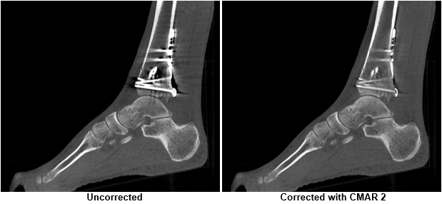
Figure 1. Sagittal plane through ankle. Dark and bright streaking on left image has been greatly reduced.
Figure 2 demonstrates the effectiveness of metal artifact reduction software when applied to a patient with internal fixation of the radius with a plate and screws. The blooming effect has been reduced, allowing better visualization of the fracture as well as the metal-bone interface along the radius.
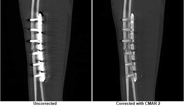
Figure 2. Sagittal plane through arm, showing internal fixation of the radius with plate and screws. Radius fracture is better visualized after metal artifact reduction is applied to reduce metal blooming.
Figure 3 below demonstrates reconstruction of the lower leg with a tibial shaft fracture fixated with an intramedullary rod. Metal artifacts that propagated through the soft tissue have been reduced and the metal-bone interface is better visualized.
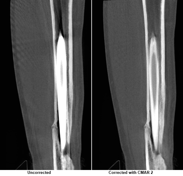
Figure 3. Sagittal plane through lower leg, showing tibial shaft fracture fixated with an intramedullary rod. Metal artifacts and dark streaking have been greatly reduced and the metal-bone interface is better visualized.
An even more challenging problem is the correction of artifacts caused by large metal implants, such as total knee arthroplasty (TKA) shown in Figure 4. In this example, the application of metal artifact reduction software has allowed better visualization of the metal-tissue interface. This can aid in the evaluation of tissue healing in the vicinity of the implant.
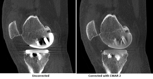
Figure 4. Sagittal plane through knee, showing TKA hardware. Artifacts through the metal-bone interface have been reduced, improving visualization of tissue healing in the vicinity of metal implant.
Conclusion
A compact, affordable 3D extremity imaging system offers important diagnostic advantages to orthopaedic and sports medicine practices while simultaneously providing patients with greater convenience and comfort. The metal artifact reduction software can help reduce artifacts generated by highly-attenuating implants. This allows better visualization of patient anatomy as well as tissue healing in the vicinity of metal.
###
*Regulatory requirements regarding shielding may vary, depending on region.
The views, opinions and positions expressed within these guest posts are those of the author alone and do not represent those of Becker's Hospital Review/Becker's Healthcare. The accuracy, completeness and validity of any statements made within this article are not guaranteed. We accept no liability for any errors, omissions or representations. The copyright of this content belongs to the author and any liability with regards to infringement of intellectual property rights remains with them.