L&K Spine Announces World’s First Clinical Use of BluEX-LT Curved Expandable Lateral Cage
L&K Spine, the U.S. subsidiary of L&K Biomed, is proud to announce the successful first-in-human clinical use of the BluEX-LT, its state-of-the-art curved expandable lateral cage. This landmark procedure was performed by Dr. Gary D. Fleischer, a prominent orthopedic spine surgeon based in Raleigh, North Carolina.
The surgery marked a significant milestone in minimally invasive spine surgery (MISS) and adult spinal deformity correction. Dr. Fleischer successfully completed a complex 3-level (L2-L5) lumbar interbody fusion using a single incision, leveraging the synergy of L&K Spine’s BluEX-LT implant and the proprietary Shallow Psoas Docking technique.
A Staged Approach Demonstrating Unprecedented Deformity Correction
Notably, this case highlighted the implant’s profound impact on surgical planning for complex deformity patients. Dr. Fleischer utilizes a sophisticated staged approach for deformity correction: inserting the lateral cages first, obtaining standing X-rays between stages to assess real-time alignment, and then finalizing the boundaries for posterior screw fixation.
Prior to the procedure, the patient’s severe deformity indicated a more extensive, long-segment fusion with fixation spanning from T10 to S1. However, following the initial insertion and expansion of the BluEX-LT, standing imaging revealed such exceptional lordotic restoration and structural correction that Dr. Fleischer was able to dramatically downsize the posterior construct, stopping at L2 instead of T10. By finalizing the surgery as an L2-S1 fixation, multiple thoracic motion segments were preserved, significantly reducing surgical invasiveness and optimizing patient recovery.
A Curved Solution for Lateral Anatomy
The BluEX-LT is engineered as a curved solution specifically for lateral spine surgery, designed to optimize the restoration of lordosis and spinal alignment. Unlike traditional rectangular lateral cages, the BluEX-LT’s curved geometry allows it to seat accurately on the apophyseal ring to minimize subsidence and maximize deformity correction.
Key features of the BluEX-LT include:
- Stable Expansion: The vertical guide design allows for stable expansion of up to 4mm.
- Enhanced Fusion Support: The device features a post-bone packing slot, allowing for the addition of bone graft even after the cage has been expanded.
- Osseointegration: The surface is treated to facilitate osseointegration.
- Versatile Sizing: Available in multiple lengths and lordotic angles.
Synergy with Shallow Psoas Docking Technique
The success of the 3-level procedure was further enhanced by the Shallow Psoas Docking technique, a direct visualization lateral system. This approach offers several critical advantages:
- Direct Visualization: The system provides a direct view of the psoas muscle and crossing nerves, adhering to a “what you see is what you get” philosophy.
- Muscle-Sparing: It utilizes a muscle-sparing split in line with the fibers.
- Reduced Neurological Risk: By identifying and protecting nerves visually, the technique addresses concerns regarding neurological sequelae common in traditional LLIF.
- Clear Imaging: The minimalistic, radiolucent tubular retractor allows for clear fluoroscopic visualization.
- Multi-Level Efficiency: The tube-based system makes it easy to “slide” along the psoas to move from disc level to disc level through a single approach, sparing soft tissues and saving operative time.
Executive Perspective
“The introduction of the BluEX-LT represents a true paradigm shift in the treatment of spinal deformity,” said Alex Kang, CEO of L&K Spine. “By seamlessly marrying a curved anatomical footprint with advanced expandable technology, we are proud to deliver an innovative solution that establishes a new direction for minimally invasive spine surgery. Our goal is to continually provide surgeons with revolutionary tools that not only optimize complex surgical workflows but, most importantly, elevate the standard of patient care.”
Clinical Perspective
“The unique design of the BluEX-LT cage provides significant advantages over the current, prevalent rectangular cage designs. The curved endplates create a force distribution that mirrors the apophyseal ring and allows the surgeon to place forces over the cortical vertebral cylinder, minimizing the transverse concentration of forces on the unsupported endplate that predisposes the cage to subsidence. Additionally, the natural apex that is created by lordotically elevating the curved surface acts to help center the implant and makes it much more stable in resting lateral migration.
The instrumentation associated with the cage facilitates efficient insertion and expansion of the cage without displacing it with unwanted forces seen in many other systems as a side effect to engage the expansion mechanism. Post-deployment graft filling was easily facilitated through the inserter making it automated and adding no difficult “fiddle factor”. The excellent combination of this innovative and unique evolution of lateral, expandable cages, and the well thought out instrumentation greatly aided me in the care of this patient and in achieving the desired outcomes with the least risk.”
— Gary D. Fleischer, MD
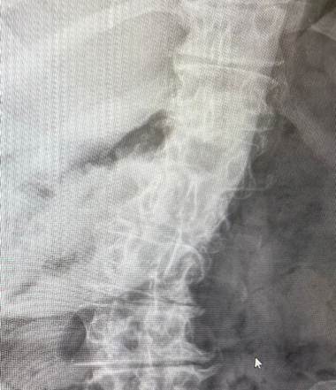
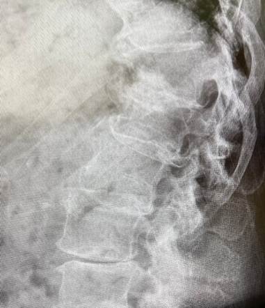
Preop Image – AP Preop Image – Lateral
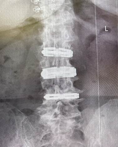
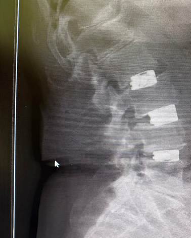
Postop Image – AP Postop Image – Lateral
At the Becker’s 32nd Annual Meeting: The Business and Operations of ASCs, taking place October 29-31 in Chicago, ASC leaders, surgeons and healthcare executives will explore strategies to drive growth, enhance operational performance, navigate reimbursement challenges and prepare for the future of ambulatory surgery. Apply for complimentary registration now.