Cobb in 1948 stated that the causative mechanism of about 90% of scoliosis is idiopathic1.
Modern researchers have identified a multitude of different factors, both biological as well as mechanical, related to either the cause or the progression of idiopathic scoliosis. There is currently no universally accepted scientific theory as to causation or progression of AIS.
This brief post highlights a clinical phenomenon observed in AIS patients and discusses a possible relationship to AIS etiology. A suggested therapy is described.
Roth postulated an interrelation between the growth of the axial skeleton and the nervous system (Roth, 1968)2.
“The growth in length of the spine proceeds in a cranio-caudal direction. The elongation of the spine is associated with the physiological growth differential between spine and nervous system which results in the ascent of the spinal cord and formation of the cauda equina.“
Roth observed that X-rays of AIS patients consistently showed a thinning and reduced height of the concave side pedicles.
At the earliest stages of embryonic development, the spinal cord fills the entire length of the spinal canal. As the physiologic growth of the vertebral column begins, the slower growing cord is tractioned upwards. These forces influence the position of the spinal cord within the spinal canal as well as the shaping of the vertebral foramina.
During periods of rapid growth, the lengthening of the spine must be accompanied by corresponding lengthening of the spinal cord and nerve roots. Roth postulates that if the neural tissues don’t keep up with the growing spine, an adaptive scoliotic curve results. Correction of the disturbed equilibrium of growth between spine and cord is achieved by lateral flexion of the spine towards the side of increased tension.
At first this lateral flexion is functional, but after maximal movement of the intervertebral joints and discs has been exceeded, structural changes of the vertebral bodies will appear.
The growth spurt of the vertebral column is associated with an increased output of hypophyseal growth hormone with stimulates the growth of all tissues except the nervous tissue.
This theory of asynchronous growth explains why AIS is progressive during rapid growth spurts. Large curves above 30-50 degrees can remain progressive after growth is complete due to mechanical forces of gravity on an imbalanced spine.
This also explains why the spinal cord adapts the shortest route along the concave side of the spinal canal.
A short cord is reflected in the common deformity of the vertebral foramen and thinning of the pedicle on the concave side.
The clinical course of the idiopathic scoliosis depends on the degree of growth difference between the spinal column and the spinal cord. The greater the difference, the more severe the scoliosis.
Porter (2001) states that the putative impaired growth of the spinal cord in AIS may result from an abnormal response to stretch. Five possible reasons are given3.
Some questions:
If the theory is correct, why is there an absence of any neurological deficit after correction of the scoliosis with bracing or surgery? If the cord is really short, wouldn’t lengthening of the spinal column cause additional stretching and damage to the neural structures? Straightening of a curved structure does not require any lengthening of the spinal cord even though sitting height can be increased. Why don’t all children with a tethered cord develop scoliosis? How does this phenomenon effect lumbar AIS? Why is neck flexion not limited in AIS? For a full list of these controversial observations see Chu 20084.
Abnormal tension in the neural tissues can be evaluated by placing the patient in a position of extension of the spine and performing the straight leg raise. If there is abnormal tension, the test will be painful for the patient and they will attempt to mitigate the tension by either flexing the knee or the lumbar spine. The patient will attempt to avoid the stretch sensation (pain) by pelvic rotation. This is seen when the patient lifts the ipsilateral hip off the examination table at the extent of their SLR test.
Straight Leg Raise (SLR) test increases neuraxial and meningeal tension due to the continuity of the PNS to the CNS5,6 . Brieg (1960) says that changes in relative lengths of the spinal canal and cord, can lead to pathologic axial tension and damage the cord and spinal nerves by overstretching7.
Passive cervical flexion, increasing lumbar lordosis, hip adduction, hip medial rotation, along with flexing the foot on the leg tensions the spinal cord to a degree and therefore potentiates the results seen in the SLR test.
The normal range of the SLR varies widely. Troup (1986) describes between 50 - 120 degrees8. The degree measurement is less important than the patient’s presentation (attempts to avoid a painful stretching sensation) and the location and quality of the pain produced at the endpoint of the test.
In his book, Mobilisation of the Nervous System, David Butler discusses the principles involved. This section of his book is well worth reading to get the finer points involved in successful application of the technique9.
Treatment time is critical to success with this technique and therefore the patient must be instructed to apply this therapy at home.
Because we are often working with a young child, it is preferable to instruct a parent on effective use of the technique.
Perform this stretching on a firm surface, the floor is preferable. Try using an open doorway as assistance if the parent cannot provide continuous support during the prolonged stretch.
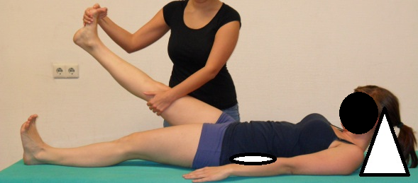
● The head should be flexed with the chin to the chest.
● A roll should be placed under the lower back, just above the belt line.
● Make sure the knee is completely straight and the foot flexed toward the head.
● Elevate the knee to a level that is 5/10 on the pain scale and hold that position for 2 minutes.
● Repeat with the opposite leg.
Pins and needles are quite common during this type of stretching. To get an effective stretch of the tissues involved, you may need to see such a neural response. If you are creating numbness in the feet in an adolescent or child, be cautious not to push past that point Typically this numbness is temporary, and evidence of the tight, in-elastic nervous system. Proceed with caution, the numbness should fully recover during the rest cycle.
About the Author:
Dr. Andrew J Strauss, BS, MS, DC, has over 30 years experience with postural and structural distortions. Beyond his Bachelor of Science and Doctorate in Chiropractic, Dr. Strauss has a Masters Degree in Acupuncture, a Graduate Diploma in clinical nutrition, is a lifetime yoga practitioner and yoga instructor, is certified in electro diagnosis (EMG/NCV) and is the Vice President of the CLEAR Scoliosis Institute.
Dr. Strauss’s practice, Hudson Valley Scoliosis Correction Center, provides non-surgical treatment for all types of scoliosis (from mild to severe). Using custom designed scoliosis-specific exercise programs, patients ranging from children to adults are able to avoid scoliosis surgery and experience rapid improvements to their curve, posture and pain. In selected cases of both adults and children, the latest in 3D scoliosis brace designs are also used as a component of the program of care.
References
Bohannon RW: Cinematographic analysis of the passive straight-leg-raising test for hamstring muscle length. Physical Therapy. 1982;62:74-1269.
Breig A: Adverse Mechanical Tension in the Central Nervous System: An Analysis of Cause and Effect. Stockholm: Almqvist & Wiksell; 1978.
Breig A: Biomechanics of the central nervous system. Stockholm: Almqvist & Wiksell; 1960.
Butler D: Mobilisation of the Nervous System. Churchill, Livingstone, Edinburgh; 1991.
Chu WC, Lam WM, Ng BK, et al: Relative shortening and functional tethering of spinal cord in adolescent scoliosis—result of asynchronous neuro-osseous growth, summary of an electronic focus group debate of the IBSE. Scoliosis. 2008;3:8.
Cobb JR: Outline for the study of scoliosis. AAOS, Instructional Course Lectures. Edited by: Edwards JW. 1948, Ann Arbor:The American Academy of Orthopaedic Surgeons, 5: 261-75.
Cox JM: Low Back Pain, Mechanism, Diagnosis, Treatment, 6th Edition. Philadelphia: Lippincott, Williams and Wilkins 1999: pgs 438-439.
Porter RW: The pathogenesis of idiopathic scoliosis: uncoupled neuro-osseous growth? Eur Spine J. 2001;10:473–481.
Roth M: Idiopathic scoliosis caused by a short spinal cord. Acta Radiol Diagn (Stockh) 1968;7:257–71.
Roth M: Idiopathic scoliosis from the point of view of the neuroradiologist. Neuroradiology. 1981;21:133–138 28.
Troup JDG, Hood CA, Chapman AE. Measurements of the sagittal mobility of the lumbar spine and hips. Part B. Annals of Physical Medicine. 1968;9:308–321.
The views, opinions and positions expressed within these guest posts are those of the author alone and do not represent those of Becker's Hospital Review/Becker's Healthcare. The accuracy, completeness and validity of any statements made within this article are not guaranteed. We accept no liability for any errors, omissions or representations. The copyright of this content belongs to the author and any liability with regards to infringement of intellectual property rights remains with them.